






















Advantages and Differences from Conventional PET Scan
Learn the advantages of digital pet radiography, how it differs from PET scans, and when veterinarians may recommend X-rays for your pet.


Diagnostic imaging helps veterinarians identify injuries, investigate illnesses, and assess changes inside a pet's body. Digital pet radiography, commonly known as digital X-rays, is one of the most frequently used imaging methods in veterinary practice. It allows veterinarians to examine bones, the chest, the abdomen, and certain dental structures without surgery.
Understanding Digital Pet Radiography can help pet owners learn how X-rays support diagnosis and why a veterinarian may recommend them instead of another imaging test. However, digital radiography and PET scans are different technologies. A PET scan, or positron emission tomography scan, uses a radioactive tracer to assess biological activity, while an X-ray primarily shows the structure and appearance of tissues.
What Is Digital Pet Radiography?
Digital pet radiography is an imaging technique that uses X-rays and an electronic detector to create images of a pet's internal structures. Instead of recording the image on traditional photographic film, the system captures the information digitally and displays it on a computer screen.
Veterinarians commonly use digital X-rays to investigate broken bones, joint abnormalities, chest conditions, swallowed foreign objects, and changes in the size or position of internal organs. The images can help identify abnormalities that require treatment or further testing.
Digital radiography is widely used because it is generally quick, accessible, and suitable for many routine diagnostic situations. Depending on the area being examined and the pet's temperament, positioning may be possible without sedation. Some animals need sedation to remain still and obtain clear images.
What Is a Conventional PET Scan?
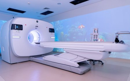
A PET scan is a nuclear medicine imaging technique that evaluates biological processes inside the body. It involves administering a small amount of a radioactive tracer, which accumulates in tissues according to the characteristics of the tracer and the biological process being investigated.
The scanner detects signals from the tracer and uses them to create images showing patterns of activity within the body. PET imaging is used in human medicine for selected conditions, including certain cancers and neurological disorders. Its use in veterinary medicine is more specialised and less widely available than routine X-ray imaging.
PET scans and digital X-rays serve different purposes. An X-ray shows structural details, while a PET scan provides information about activity within tissues. In some clinical settings, PET imaging may be combined with computed tomography (CT) to relate functional information to anatomical structures.
Key Differences Between Digital Pet Radiography and PET Scans
Although both technologies help investigate medical conditions, they differ in how they produce images, what they reveal, and when they are useful.
1. How the Images Are Produced
Digital radiography sends a controlled beam of X-rays through the body. An electronic detector records the radiation that passes through the tissues. Differences in tissue density create the image, with bones generally appearing lighter than surrounding soft tissues.
PET imaging works differently. A radioactive tracer is introduced into the body, and the scanner detects signals produced as the tracer decays. The resulting images show how the tracer is distributed, helping clinicians assess selected biological processes.
2. What Each Imaging Method Can Show
Digital X-rays are particularly useful for examining bones, fractures, joint changes, the chest, and certain abdominal abnormalities. They can also help detect some foreign objects and assess the position or size of organs.
PET scans focus on biological activity rather than simply showing anatomical structure. Depending on the tracer and the clinical question, PET imaging can help identify areas of increased or decreased metabolic activity.
Neither method answers every diagnostic question. A veterinarian chooses the imaging test according to the suspected condition and the information needed.
3. Speed and Accessibility
Digital X-ray images are usually available within seconds after exposure. This allows the veterinary team to review the images promptly and decide whether additional views or tests are needed.
PET imaging involves additional preparation, including tracer administration and time for the tracer to distribute through the body. The scan itself and the associated procedures can take considerably longer than routine radiography.
Digital X-ray equipment is available in many veterinary clinics. PET scanning requires specialised equipment, trained personnel, and appropriate facilities for handling radioactive tracers. As a result, it is not routinely available in general veterinary practices.
4. Cost and Practical Considerations
Digital radiography is generally more affordable than specialised advanced imaging. It is commonly used as an initial diagnostic test because it can provide useful information without the expense and complexity of a nuclear medicine procedure.
PET scans typically involve higher costs and more specialised resources. They are considered when the clinical question requires functional imaging and when the expected diagnostic benefit justifies the procedure.
Actual costs depend on the veterinary facility, the body region being examined, the number of images required, and whether additional procedures or tests are necessary.
Advantages of Digital Pet Radiography
Digital radiography offers several practical benefits for veterinary diagnosis.
Quick Image Availability
One of the main advantages is the speed at which images can be reviewed. The veterinary team can assess the results shortly after taking the X-rays and determine whether additional views are necessary.
Convenient Image Storage
Digital images can be stored in the pet's medical record and retrieved during future appointments. Comparing earlier and newer images may help veterinarians assess changes in fractures, joint conditions, or other structural abnormalities.
Image Adjustment and Sharing
Veterinarians can enlarge images and adjust display settings to examine particular areas more closely. Digital files can also be shared with veterinary radiologists or referral specialists when a second opinion is needed.
Useful for Common Veterinary Conditions
Digital X-rays can help investigate a wide range of problems, including suspected fractures, arthritis, chest abnormalities, and certain digestive tract concerns. They are often an appropriate first step when the veterinarian suspects a structural problem.
No Radioactive Tracer Required
Unlike PET imaging, digital radiography does not require the administration of a radioactive tracer. It uses an external X-ray source to produce the image.
X-rays still involve ionising radiation, so veterinary teams use appropriate exposure settings and radiation safety procedures. The benefits and risks of imaging are considered for each patient.
Are There Any Limitations to Digital Pet Radiography?
Digital X-rays provide valuable information, but they do not show every condition clearly. Because radiographs produce two-dimensional images, structures may overlap, making some abnormalities difficult to assess.
Soft tissue changes can also be challenging to distinguish when different tissues have similar X-ray appearances. For certain conditions, the veterinarian may recommend ultrasound, CT, MRI, laboratory testing, or another diagnostic procedure.
PET scans have different limitations. They require specialised equipment, involve radioactive tracers, and may not be appropriate for every condition. Functional imaging findings may also need to be interpreted alongside anatomical imaging and other diagnostic results.
The most advanced or expensive test is not automatically the best choice. The appropriate method depends on the suspected disease, the pet's condition, and the clinical information needed to guide treatment.
When Might a Veterinarian Recommend Digital X-Rays?
A veterinarian may recommend digital radiography when a pet has persistent limping, suspected bone injury, breathing difficulties, possible ingestion of a foreign object, or signs of a condition affecting the chest or abdomen.
For example, if a dog develops sudden lameness after an injury, X-rays may help identify a fracture or joint abnormality. If a cat has respiratory symptoms, chest radiographs may help the veterinarian assess the lungs and surrounding structures.
If the X-ray findings do not fully explain the symptoms, further testing may be recommended. The decision to proceed with additional imaging depends on the examination findings and the suspected cause of the problem.
Choosing the Right Imaging Method for Your Pet
Digital pet radiography and PET scans are not interchangeable. Digital X-rays are commonly used to examine anatomical structures and investigate many routine veterinary concerns. PET scans provide specialised information about biological activity and are used in more specific diagnostic situations.
For most common injuries and structural abnormalities, digital radiography is a practical starting point. When a condition requires more detailed anatomical or functional information, the veterinarian may recommend another imaging method or a combination of tests.
If your pet is showing signs of pain, limping, breathing changes, or other unexplained symptoms, consult your veterinarian. A clinical examination will help determine whether X-rays or another diagnostic procedure is appropriate. Choosing the right test can help the veterinary team identify the problem and develop a suitable treatment plan.
What's Your Reaction?
 Like
0
 Dislike
0
 Love
0
 Funny
0
 Angry
0
 Sad
0
 Wow
0